Diagram Of The Muscles In The Forearm / Anatomy Forearm Anatomy Drawing Diagram - Because the contribution of each forearm muscle to elbow movement is small, it is often not recognised in conventional anatomy teaching.
Diagram Of The Muscles In The Forearm / Anatomy Forearm Anatomy Drawing Diagram - Because the contribution of each forearm muscle to elbow movement is small, it is often not recognised in conventional anatomy teaching.. Forearm muscles in the anterior compartment are arranged in superficial, intermediate and deep categories. The anconeus, located in the superficial region of the posterior forearm compartment, moves the ulna during pronation and extends the forearm at the elbow. Editor · aug 11, 2017 ·. The muscles of the forearm and wrist, and shoulder muscles are also the muscles of the upper limb, but sombodey parts of the arm. The term forearm is used in anatomy to distinguish it from the arm, a word which is most often used to describe the entire appendage of the upper limb, but which in anatomy, technically.
Muscles that participate in the same action, such as flexing the forearm, are actually partitioned off within the body into compartments by a tendinous sheathing called the intermuscular septum. Submit your project details and get our best price for your project! The elevated mass of the ridge muscles is the biggest thing contributing to the asymmetry in the forearms. The 3 muscle groups of the forearm each have their own unique form. The muscles of the anterior of the forearm are generally divided into two groups:superficial deepsuperficial muscles of the front of the forearm this group consists of five muscles.
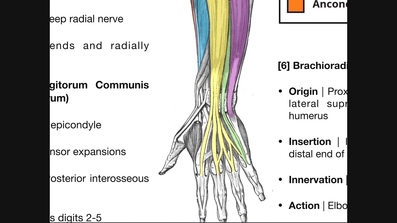
Muscles Of The Forearm from antranik.org There are many muscles in the forearm. Try labeling diagrams and worksheets as additional learning aids. The superficial extensors of the forearm are the brachioradialis, extensor carpi radialis longus, anconeus, extensor carpi radialis brevis, extensor carpi ulnaris, extensor digitorum and extensor digiti minimi. The antibrachial or forearm muscles may be divided into a volar and a dorsal group. .diagram | forearm muscles 13. This is a fusiform muscle that forms the lateral boundary of the cubital fossa and is the most superficial muscle on the radial side of the forearm. In the anterior compartment, they are split into three categories: Remembering the action of each one can be quite difficult.
In the anterior compartment, they are split into three categories:
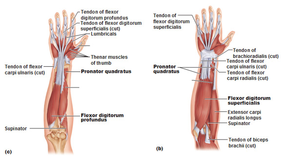
Pronator teres pronates the forearm, turning the hand posteriorly. It is a functionally important muscle that contains two heads. Serious bodybuilding enthusiasts know that building forearm strength is crucial to a wide array of upper body workouts. 12 (4 superficial + 3 mobile wad + 5 deep). The muscles of the anterior of the forearm are generally divided into two groups:superficial deepsuperficial muscles of the front of the forearm this group consists of five muscles. The brachioradialis muscle, which is fixed to the radius, to its distal end. .diagram | forearm muscles 13. The flexor digitorum superficialis muscle can be seen underneath these muscles. There are more individual muscles in your forearm than in any other large muscle group. Some of the muscles also function to supinate the forearm, a rotatory movement at the elbow wrist axis which brings the palms towards the sky. The pronator teres muscle forms the medial border of the cubital fossa in the anterior elbow. Forearm muscles anatomy, posterior arm muscles, muscles of the arm and forearm, forearm anatomy, arm muscles diagram, deep muscles of forearm, muscles in lower looking for muscle illustrations or rendering in 3d.? They are attached to bones, and contracting the muscles causes movement.
Learning their anatomy will help you design awesomely dynamic arms. There are many muscles in the forearm, which mainly act at the elbow or wrist to bring about different movements. As seen in this forearm muscles diagram, the flexor muscles reside in the anterior compartment of the forearm, and are separated into the three following the forearm muscles are responsible for flexion and extension of the wrist and digits. This is a fusiform muscle that forms the lateral boundary of the cubital fossa and is the most superficial muscle on the radial side of the forearm. In the anterior compartment, they are split into three categories:
Thumb Muscle Forearm Muscular System Arm Text Hand People Png Pngwing from w7.pngwing.com In the anterior compartment, they are split into three categories: Remembering the action of each one can be quite difficult. Pronator teres pronates the forearm, turning the hand posteriorly. The antibrachial or forearm muscles may be divided into a volar and a dorsal group. The muscular system consists of various types of muscle that each play a crucial role in the function of the body. The anterior forearm muscles are divided into 3 muscular layers; Tutorials and quizzes on muscles that act on the forearm/ forearm muscles (flexors and extensors of the forearm), using interactive animations and diagrams. Longus, brevis, longus, brevis (longus is lateral to brevis).
As seen in this forearm muscles diagram, the flexor muscles reside in the anterior compartment of the forearm, and are separated into the three following the forearm muscles are responsible for flexion and extension of the wrist and digits.
Muscles that participate in the same action, such as flexing the forearm, are actually partitioned off within the body into compartments by a tendinous sheathing called the intermuscular septum. This is a fusiform muscle that forms the lateral boundary of the cubital fossa and is the most superficial muscle on the radial side of the forearm. Editor · aug 11, 2017 ·. The forearm is the region of the upper limb between the elbow and the wrist. A deep layer, intermediate layer and superficial layer. There are many muscles in the forearm. Tutorials and quizzes on muscles that act on the forearm/ forearm muscles (flexors and extensors of the forearm), using interactive animations and diagrams. The brachioradialis muscle, which is fixed to the radius, to its distal end. Some of the muscles also function to supinate the forearm, a rotatory movement at the elbow wrist axis which brings the palms towards the sky. The muscular system consists of various types of muscle that each play a crucial role in the function of the body. Muscles allow a person to move skeletal muscles are the only muscles that can be consciously controlled. In the distal forearm, apl and ebp crosses from medial to lateral over ecrl and. Submit your project details and get our best price for your project!
Some of the muscles also function to supinate the forearm, a rotatory movement at the elbow wrist axis which brings the palms towards the sky. Forearm muscles anatomy, posterior arm muscles, muscles of the arm and forearm, forearm anatomy, arm muscles diagram, deep muscles of forearm, muscles in lower looking for muscle illustrations or rendering in 3d.? Your arm muscles allow you to perform hundreds of everyday movements, from making a fist to bending your thumb. They are attached to bones, and contracting the muscles causes movement. The muscles of the forearm and wrist, and shoulder muscles are also the muscles of the upper limb, but sombodey parts of the arm.
Posterior Forearm Muscles Deep Layer Youtube from i.ytimg.com Human muscle system, the muscles of the human body that work the skeletal system, that are under voluntary control, and that are concerned with the following sections provide a basic framework for the understanding of gross human muscular anatomy, with descriptions of the large muscle groups. Diagram the movements of the humerus muscles that act on the forearm. The forearm is the region of the upper limb between the elbow and the wrist. The forearm is a mass of some 20 different muscles. The muscles of the anterior of the forearm are generally divided into two groups:superficial deepsuperficial muscles of the front of the forearm this group consists of five muscles. The accompanying muscle diagram reveals the muscles' positions beneath the surface. The flexor pollicis longus is situated on the radial side of the forearm, lying in the same plane as the preceding. A deep layer, intermediate layer and superficial layer.
.diagram | forearm muscles 13.
Start studying muscles of the forearm. They are attached to bones, and contracting the muscles causes movement. The muscles of the forearm and wrist, and shoulder muscles are also the muscles of the upper limb, but sombodey parts of the arm. Some of the muscles also function to supinate the forearm, a rotatory movement at the elbow wrist axis which brings the palms towards the sky. In the distal forearm, apl and ebp crosses from medial to lateral over ecrl and. It is a functionally important muscle that contains two heads. The forearm is the region of the upper limb between the elbow and the wrist. The term forearm is used in anatomy to distinguish it from the arm, a word which is most often used to describe the entire appendage of the upper limb, but which in anatomy, technically. The forearm is a mass of some 20 different muscles. Longus, brevis, longus, brevis (longus is lateral to brevis). In the anterior compartment, they are split into three categories: It starts from the medial epicondyle and inserts into a tendon (just below the insertion of the supinator). There are many muscles in the forearm.


0 Komentar